Application of Functional MRI in Parkinson’s Disease and Default Mode Network: Review of the Literature
Investigating Parkinson’s Disease Through fMRI and the Default Mode Network
We're proud to highlight new research coming out of the Brain Health and Wellness Lab ([insert website link]) and authored by Holly Bardutz, along with three of her research assistants—now all proud medical students (congratulations to the team!)—and Dr. Rehman, a neurologist with the Saskatchewan Health Authority.
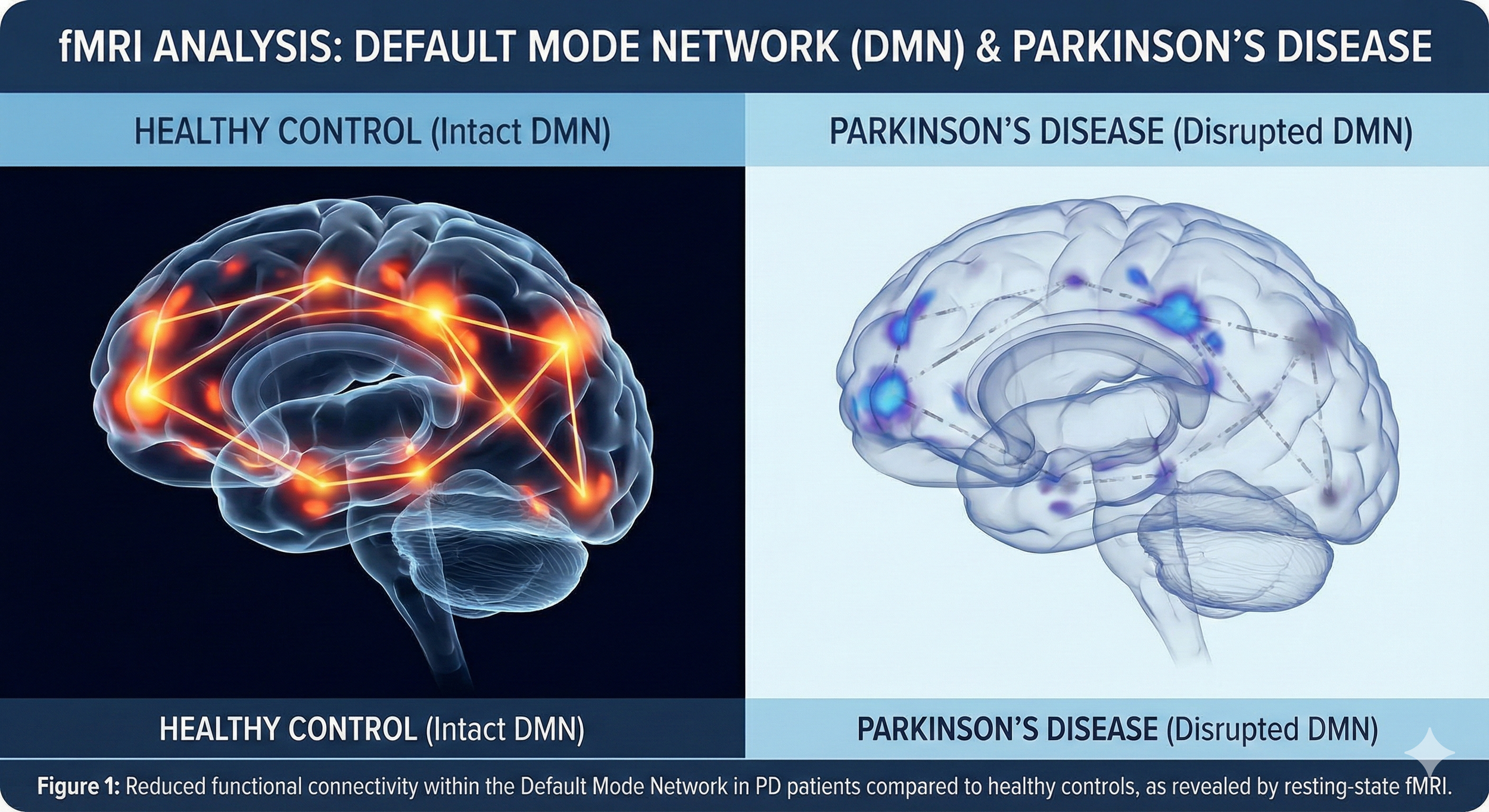
The article was recently published in the Canadian Journal of Neurological Sciences (May 2025). This comprehensive review examines how resting-state functional MRI (fMRI) reveals changes in the Default Mode Network (DMN) in individuals with Parkinson’s disease (PD).
The DMN is a key brain network involved in memory, self-reflection, and social cognition. In people with PD, researchers found disrupted connectivity in core regions of the DMN, such as the posterior cingulate cortex (PCC), medial prefrontal cortex (mPFC), and precuneus. These changes are linked not only to motor symptoms but also to cognitive decline, including challenges with memory and executive function.
The review also explores how dopamine loss, along with other neurotransmitter imbalances and neuroinflammatory processes, may contribute to these disruptions. Importantly, some studies indicate that dopaminergic treatments may partially restore DMN function, offering insight into how fMRI could serve as a biomarker to track disease progression and treatment response.
Another key takeaway is the variability in DMN connectivity across PD subtypes, which points to the importance of personalized approaches in both research and care.
Why It Matters: This research strengthens the case for using advanced imaging tools to better understand the full scope of Parkinson’s, beyond movement symptoms. It opens up new avenues for diagnosis, monitoring, and individualized interventions in neurological health.